Acute central nervous system catastrophes and extreme environmental exposures produce profound neurohormonal surges that manifest as dramatic electrocardiographic abnormalities mimicking acute myocardial infarction.
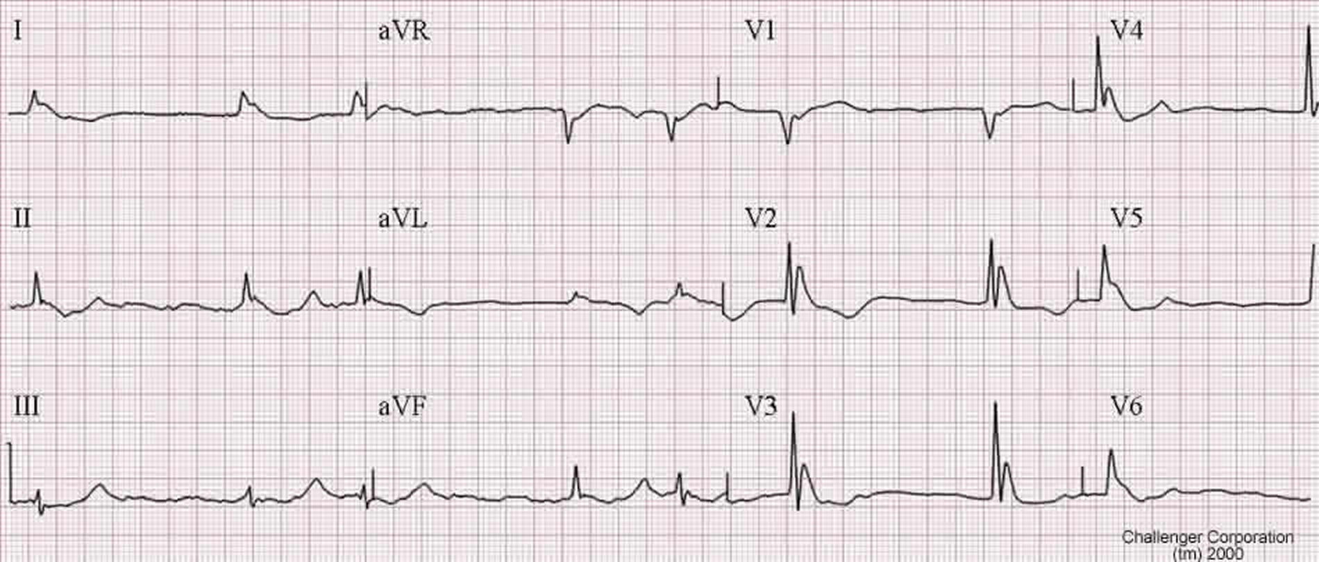 Figure 19.1: 12-lead ECG in severe Hypothermia showing prominent Osborn (J) waves at the J-point, baseline somatic muscle tremor artifact, and marked bradycardia. Rendered with solid white background.
1. Intracranial Hemorrhage & Neurogenic "Cerebral T Waves"
- Pathophysiology: Massive intracranial pressure elevation (Subarachnoid Hemorrhage [SAH], severe traumatic brain injury) triggers a massive sympathetic hypothalamic-medullary surge, releasing excessive norepinephrine directly into myocardial nerve terminals and causing focal myocyte necrosis (contraction band necrosis / neurogenic stunned myocardium).
- Electrocardiographic Hallmarks:
- "Cerebral T Waves": Enormous, wide, deeply inverted, symmetrical T waves ($>10-15\text{ mm}$ amplitude) across precordial leads ($V_1 - V_6$) and limb leads.
- Extreme $QTc$ Prolongation: Frequently exceeding $600-700\text{ ms}$.
- Prominent U waves and sinus bradycardia.
- Transient ST-segment elevation or depression mimicking acute STEMI.
2. Hypothermia & The Osborn (J) Wave
- Pathophysiology: Severe core hypothermia ($<32^\circ\text{C}$) exaggerates the transmural voltage gradient by selectively prolonging epicardial $I_{to}$-mediated action potential notches relative to the endocardium.
- Electrocardiographic Triad:
- Osborn Wave (J Wave): Prominent positive dome-shaped deflection at the junction of the QRS complex and ST segment (height is directly proportional to the severity of hypothermia).
- Baseline Somatic Tremor: High-frequency shivering muscle artifact.
- Progressive Conduction Slowing: Sinus bradycardia $\to$ PR/QRS/QT prolongation $\to$ slow atrial fibrillation $\to$ spontaneous Ventricular Fibrillation ($<28^\circ\text{C}$) or asystole.
3. Evidence-Based Emergency Resuscitation Protocols
- Severe Hypothermia Resuscitation:
- Continuous core temperature monitoring (esophageal/bladder).
- Active Core Rewarming: Warmed IV fluids ($40-42^\circ\text{C}$), humidified heated oxygen, bladder/peritoneal lavage, or Extracorporeal Life Support (ECLS / ECMO).
- In ventricular fibrillation at core temp $<30^\circ\text{C}$: Attempt initial defibrillation up to 3 shocks; defer further shocks and medications (epinephrine) until core temp $>30^\circ\text{C}$ due to impaired hepatic/renal drug clearance.